Capillary leakage important in COVID-19 respiratory distress
by Dr. Liji Thomas, MDThe pandemic of COVID-19 caused by the severe acute respiratory syndrome coronavirus 2 (SARS-CoV-2) has now spread to over 188 countries and territories, with over 5.4 million cases, leaving more than 344,000 dead. The chief cause of death is acute severe respiratory failure.
Now, a new study published on the preprint server medRxiv* reports the pivotal role played by protein leakage through lung capillaries and promotes the use of serum albumin as a biomarker of disease progression and severity.
Lung Disease in COVID-19
COVID-19 patients typically begin with mild cold-like symptoms, which progress from the early viral response phase through the lung phase to the hyperinflammation phase. The most severe phase of the disease is characterized by progressive respiratory failure with a lack of oxygenation, culminating in acute respiratory distress syndrome (ARDS).
Hyperimmune Response
This progression is thought by some scientists to be due to a hyperimmune response to the viral infection. This is characterized by elevated levels of proinflammatory cytokines and chemokines, which both signal the recruitment of more immune cells and the activation of inflammatory pathways. These cytokines include IL-1β, IL-6, and TNF-α.
The end result of this process is severe injury to the lung tissue as well as to multiple other organs. The most significant impact is on sites that have a high expression of ACE2, the host cell receptor for SARS-CoV-2, which produces the clinical feature of what is called viral sepsis.

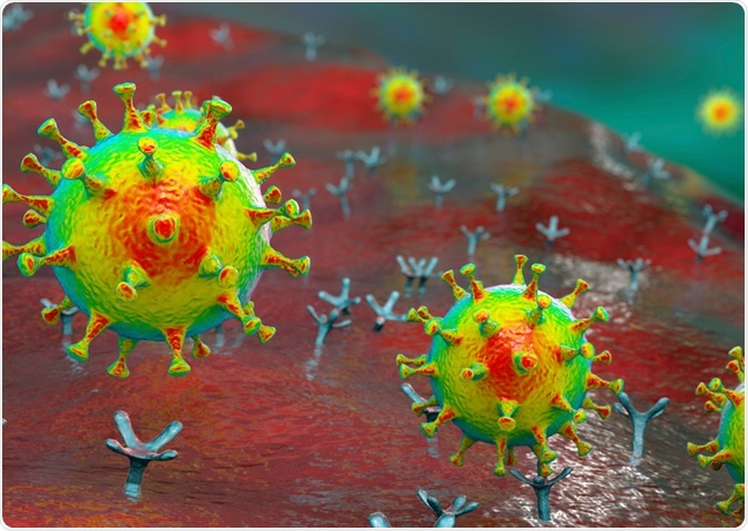
SARS-CoV-2 viruses binding to ACE-2 receptors on a human cell, the initial stage of COVID-19 infection, conceptual 3D illustration. Credit: Kateryna Kon / Shutterstock
Direct Viral Insult
Electron microscopy shows the direct damage caused by the viral particles as well, confirmed by the presence of viruses in the bronchial cells and type II alveolar pneumocytes.
Endothelial Dysfunction
Another mechanism of injury is endothelial dysfunction. This is the result of the endothelial cell reaction to the invading virus, hypoxia, immune activation, and the effect of inflammatory mediators, including cell damage that causes histone release. In this way, there are multiple sources of endothelial damage, that increase the odds of inflammation, increased coagulation, and vasoconstriction.
The endothelial dysfunction occurring in many different organs causes both endothelial and immune cells to undergo programmed cell death or apoptosis. This may cause a breakdown of the epithelial-endothelial barrier, resulting in the presence of exudate in the alveolar cavity.
Chest Imaging Supports Exudative Edema
This mechanism is reflected in chest imaging results. The B lines, white lung and patchy pattern on lung ultrasound, as well as the ground glass opacities and slightly more dense hazy areas on chest CT, are the result of interstitial and alveolar lesions. Scientists have been looking into the reason for this alveolar exudate.
How Did the Study Examine Hypoalbuminemia in COVID-19?
The current study focuses on the presence of markedly reduced albumin levels in symptomatic COVID-19 patients, which could not be explained by the presence of protein loss in urine or the gut. Moreover, albumin infusion failed to correct the deficit.
The study was carried out on all patients aged 18 years and above, with lung disease due to COVID-19 confirmed by RT-PCR, from throat swab samples, and in whom serum albumin was measured within 72 hours of admission, in a single hospital in Milan, Italy. The study period was from February 21 to April 15, 2020.
Patients who failed to sustain respiratory function with helmet-mediated continuous positive airway pressure (CPAP) were admitted to the intensive care unit (ICU)
How Was Hypoalbuminemia Related To COVID-19 Lung Disease?
There was no significant difference in demographic and medical characteristics between the different groups in the trial. Patients in the ICU were more likely to be male, while intermediate medical ward (IMW) inpatients were more often smokers, or had a history of heart disease, stroke, or diabetes.
The symptom onset to admission duration was shorter in ICU patients at about six days, on average, vs. eight days for medical ward admissions. However, there was no difference in the time from admission to the initiation of CPAP. Nonetheless, the initial positive end-expiratory pressure was higher at this point in ICU vs. IMW patients.
ICU patients stayed in hospital for an average of 20 days compared to 8 days for IMW patients, and they also had more than double the mortality compared to IMW patients ((52.4% vs. 21.7%). This trend also held good when patients were compared based on the median albumin level, 24 g/L, at survival rates of 53.2% vs. 21%.
Serum biomarkers of inflammation and organ dysfunction were higher in ICU patients, but lymphocytes counts were reduced markedly in this group.
Albumin Levels and Outcome
In all patients, mean serum albumin levels were low, but more markedly in ICU than in IMW patients, at 20 vs. 28 g/L. The greatest negative impact on respiratory function was in ICU patients.
Within 48 hours of admission, the spot protein measurements for 77 ICU patients were undetectable in 95%. The median quantitative proteinuria measurement for 12 ICU patients was 76 mg. Among all patients, only one had a history of any condition that could account for such massive protein loss.
The investigators then classified the patients into three subgroups based on either the P/F ratio or the Brixia score on chest X-ray. They found that the worse the respiratory function, the lower was the albumin level, on average.
The three P/F categories were > 250, 250-150, and <150, with corresponding median albumin levels of 31, 24, and 20 g/L, respectively.
The falling albumin levels were linked to rising levels of IL-8, which is inflammatory in function, and inversely proportional to the anti-inflammatory IL-10.
When the chest X-ray showed more extensive and marked lung changes, the Brixia scores <5, 6-11, and ≥ 12 were associated with albumin levels of 31, 27, and 21 g/L, respectively. The corresponding microscopic tissue changes in the lung tissue included loss of alveolar membranes and the deposition of hyaline membranes in the alveoli. In addition, the interstitial tissue showed patchy swelling by a mild proliferation of fibrous tissue, edema, and lymphocyte invasion to a moderate degree.
Electron microscopy revealed that junctional complexes that are mostly responsible for the epithelial integrity were strikingly disrupted in 80% of examined samples (8/10 patients), with surfactant loss in type 2 pneumocytes. Viral particles were also present in the cytoplasm of these cells and the endothelium.
The Implications for Future Practice
The conclusion is inescapable that the hypoalbuminemia linked to COVID-19 lung disease is commensurate with the severity of respiratory distress.
The scientists theorize that the virus causes pulmonary capillary leakage mediated by the intense inflammatory reaction, which is a major cause of the disease manifestations as well as the laboratory and imaging results.
A reduction of serum albumin is frequently observed in inflammation and acute illness. The reasons include increases in the permeability of blood vessels, with albumin entering extravascular spaces more freely while it is more readily broken down.
Even though the synthesis of albumin is increased in such states, including trauma, infection or shock, the intracellular breakdown of albumin results in a lower total mass, and this indicates a poorer outcome as well as a negative response to a second insult, such as surgery. On the other hand, albumin administration systemically is not consistently linked to a better outcome in such patients, except in the case of septic shock.
In acute influenza A and other viral pneumonias, the virus directly injures the epithelium-endothelial barrier causing exudative edema, with rich protein content. Endothelial cells form almost a third of all cell types in the lungs, and thus. However, their contribution to limiting protein flux is relatively limited, the summated effect is very significant.
Autopsy of patients in the current study showed that both sides of the barrier were extensively damaged, allowing increased passage of both fluid and protein from the blood to the lung alveoli. Endothelial damage may also trigger coagulation pathways, which act together with a capillary fluid loss to cause clotting within the microcirculation.
As a result of such findings, drug trials are underway to test the efficacy and safety of drugs that prevent the leakage of blood and fluids from the capillaries into the lung alveolar space or avert disruption of the epithelial barrier.
*Important Notice
medRxiv publishes preliminary scientific reports that are not peer-reviewed and, therefore, should not be regarded as conclusive, guide clinical practice/health-related behavior, or treated as established information.
Journal reference:
- Wu, M. A. (2020). COVID-19: The Key Role of Pulmonary Capillary Leakage. An Observational Cohort Study. medRxiv preprint. doi: https://doi.org/10.1101/2020.05.17.20104877. https://www.medrxiv.org/content/10.1101/2020.05.17.20104877v1